Projects
Since its advent, magnetic resonance imaging (MRI) has revolutionised the research in fundamental neuroscience. MRI is a non-irradiating and non-invasive imaging technique offering several modalities for studying the human brain from different angles, opening new perspectives previously unconceivable for studying the brain. In particular, diffusion MRI (dMRI) exploits the thermal random motion of water molecules in biological tissues for mapping the local axonal structure at each imaging voxel. By using this information, tractography algorithms estimate trajectories capturing coherent orientations of maximal diffusion that are likely to represent real white matter fibre tracts linking together distinct grey matter areas of the brain. This comprehensive map of neural connections of the brain is called “connectome”. At the macroscopic scale, the connectome can be seen as a network, where each vertex represents a well-defined cortical or sub-cortical structure and the edges quantify the structural white matter connectivity as measured with tractography.
In the following you can find some of the projects we are currently working on. Please do not hesitate tocontact us (hardi@epfl.ch) if anything is of interest or you need more information. If you are interested in an internship or in a semester/master project with us on a related topic, please feel free to contact directly one of the people in charge of each project.
Local reconstruction
People involved: Anna Auria Rasclosa, Alessandro Daducci![]() The anisotropy of diffusion can be exploited for mapping the structural neuronal connectivity and structures invisible with other imaging modalities can be highlighted. Obviously, the reconstruction quality in each voxel has a direct impact on the final result of this fiber-tracking. The study of neuronal connectivity is of major importance in a fundamental neuroscience perspective as well as in clinical applications, such as the understanding of the Stroke, Schizophrenia, or Parkinsons disease. Consequently, our ability to achieve high angular resolution represents an important challenge. Despite all the potential of diffusion MRI, its main drawback is still the long scan time, which has limited its use in clinical practice. Accelerated acquisitions, relying on a smaller number of sampling points, are thus required.
The anisotropy of diffusion can be exploited for mapping the structural neuronal connectivity and structures invisible with other imaging modalities can be highlighted. Obviously, the reconstruction quality in each voxel has a direct impact on the final result of this fiber-tracking. The study of neuronal connectivity is of major importance in a fundamental neuroscience perspective as well as in clinical applications, such as the understanding of the Stroke, Schizophrenia, or Parkinsons disease. Consequently, our ability to achieve high angular resolution represents an important challenge. Despite all the potential of diffusion MRI, its main drawback is still the long scan time, which has limited its use in clinical practice. Accelerated acquisitions, relying on a smaller number of sampling points, are thus required.
In this project, we define advanced acquisition strategies for accelerated high angular resolution diffusion MR imaging (HARDI). In each voxel of the brain, the imaging problem for diffusion is formulated in terms of compressive sampling problems for the recovery of a sparse signal on the sphere. Such optimization problems are solved by means of state-of-the-art non-linear algorithms principally in the context of convex optimization.
Global fiber-tracking
People involved: Alia Lemkaddem, Alessandro Daducci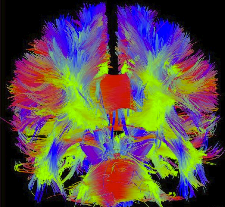 Tractography (also called fiber-tracking) is a general term for methods to reconstruct fiber pathways in the white matter based on diffusion MR imaging. It offers a unique possibility to gain insight into the structure of the human brain non invasively and in vivo. The information inferred in this manner is not only of high value for visualization of the brain connectivity and segmentation of the brain into different functional areas, it also has the potential to provide essential information that can be exploited e.g. for neurosurgical planning or for better understanding major diseases such as multiple sclerosis, epilepsy, schizophrenia, brain plasticity after strokes etc. Tractography is thus a central tool in MR-based brain connectivity analysis. Although largely developed, tractography still mostly relies on local optimization methods, and remains therefore quite unstable and unreliable, especially for mapping non-dominant pathways and when used with advanced dMRI acquisition schemes such as HARDI.
In this project, we develop new global tractography algorithms to improve significantly the accuracy and the stability of the brain connectivity analysis. Since the local methods only consider the information of one voxel at each step of the processing, this new method will try to estimate fiber pathways that are compatible with the acquired signal on a global level.
Brain networks
People involved: Alessandra Griffa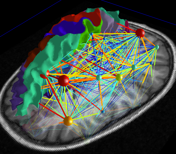 In recent years developments in graph theoretical analysis have been more and more applied to the study and characterization of both functional and structural brain networks. The human connectome has been characterized as a small-world, modular, hierarchical network, with few central nodes acting as connectors between local communities, and highly interconnected between them. This brain characterization is common to both structural and functional connectivity. Nevertheless the correlation between the two has not yet been clearly uncovered.
Our research interests include the characterization and formalism of the structure-function relationship. Particularly, we are interested in investigating the modular structure of the brain network, and the correlation between structural and functional connectivity under a hierarchical prospective.
Statistical analysis
People involved: Djalel Eddine MeskaldjiWe develop new statistical tools adapted to brain connectivity analysis, especially, the node-based or connection based inference. Such analysis involves a huge number of tests and, is related to the problem of multiple comparisons or multiple testing. The work covers two aspects.
The general problem of multiple comparison procedures. The objective is to find a multiple comparison procedure that is optimal under a certain criterion. The criterion makes the compromise between the false and the true discoveries via a weighted classification. We are now focusing on the best choice of multiple comparison procedure in the family of the scaled MCPs.
- The multiple comparison procedures under dependence. In many fields of application and particularly, in the field of brain connectivity, the observations are structured into positively dependent components. The aim is to use this information to increase the power of detecting real effects.
The developed methods are implemented in our open-source tool called Connectome Analyzer.
Clinical applications
People involved: Elda Fischi Gomez, Alessandra Griffa, Alia LemkaddemIn the following we list some of the past/ongoing clinical project conducted in our lab. The list is not exhaustive.
A Multi-Scale Registration Procedure for Automated Labeling Cortical Surfaces: Parcellating Newborn Cortex
This project is in collaboration with the Department of Development and Growth of the HUG (Hopitaux Universitaires de Genève) and its goal is to determine whether the impairment in executive functions is associate with specific regions of structural brain abnormalities in premature newborns at brith and at the age of 6 years old. Survival of newborns born extremely prematurely (EP) and born after pregnancies complicated with placental problems leading to intrauterine growth restriction (IUGR) have increased steadily due to improvement in perinatal care, though long term neurodevelopmental impairment in these survivors is increasingly recognized as a global burden to society. Although it has been shown that the organization of the brain following the preterm birth (with or without IUGR) is different from full-term born children (with appropriate birth weight) it still remains challenging to answer to which extent the ex-utero and in-utero environment influences the underlying brain structure. Neuroimaging studies on prematurely born children (with or without IUGR) have been primarily focused on the assessment of the cerebral cortex (in terms of thickness, volume and cortical surface complexity), volumes of deep gray matter structures, hippocampus and cerebellum. However, an increased interest has appeared recently towards assessing abnormalities in the development of the complex white matter (WM) fiber structure. When dealing with paediatric data, in order to map brain neuronal paths and to compare them between individuals, we first need a reliable registration between subjects and an accurate cortical surface extraction and parcellation, which implicitly means that we need an accurate tissue segmentation and gray matter - white matter surface detection. However, the major drawback in adapting ’adult procedures’ on newborn data sets is that, aside the different contrast in T1 and T2 images, almost all processing algorithms (for segmentation/registration/cortical parcellation) are based on adult atlas and templates that differ from neonate brain structures. In the frame of this work we intend to develop a reliable automatic method for newborn cortical parcellation and inter/intra-subject registration based on aligning individual’s cortical folding patterns in establish correspondence between homologous anatomical regions. In that way, we expect to understand the neurostructural origin of the long term developmental disabilities and outcome in children born prematurely and, specially, to determine if anatomical measurements at age 6 years old correlate to structural measurements at birth.
Anatomical and functional connectomes in schizophrenia and progression of psychosis
This project is in collaboration with the University Hospital of Lausanne (CHUV) and the University of Lausanne (UNIL). Schizophrenia is a disease of widespread biochemical, functional and structural brain alterations that are likely to be caused by structural and functional dys-connectivity and lead to functional impairment and positive symptoms. Redox dysregulation and impairment of glutathione (GSH) synthesis, a major antioxidant and redox buffer of the cells, have been proposed as a potential hub of the pathology, where genetic and environmental factors converge. The exact nature and pattern of the brain network alterations occurring in schizophrenia is not clear yet. Published findings are prone to a substantial variability, and this is particularly true for the early stage of psychosis. The main goals of our investigation are to identify and characterize brain connectivity modifications occurring in the early stage of psychosis, assess their relationship to genetic, evaluate their evolution in time, and investigate the link with dimensional aspects of psychosis. Both structural connectivity derived from diffusion spectrum imaging (DSI), and functional connectivity estimated from resting state fMRI are investigated. The relationship structure-function may be a potential key aspect for the characterization of neurodevelopmental brain disorders and schizophrenia.
Structural connectivity analysis of patients with epilepsy
In collaboration with the Epilepsy Unit at the HUG (Hopitaux Universitaires de Genève), we are developing advanced methods for structural connectivity analysis of epileptic patients. More precisely, we are investigating the effect of patients with temporal lobe epilepsy (MTLE) that suffer from widespread subtle white matter abnormalities and abnormal functional connectivity extending beyond the affected lobe, as revealed by Diffusion Tensor MR Imaging, volumetric and functional MRI studies. Diffusion Spectrum Imaging (DSI) is a diffusion imaging technique with high angular resolution for improving the mapping of white matter pathways. We use DSI, connectivity matrices and topological network measures to investigate how the alteration in structural connectivity influences whole brain structural networks. Furthermore, some local analysis are carried out where fiber propagation from the epileptic focus is analyzed and compared to other imaging modalities, such as Electric Source Imaging (ESI) and functional MRI.
